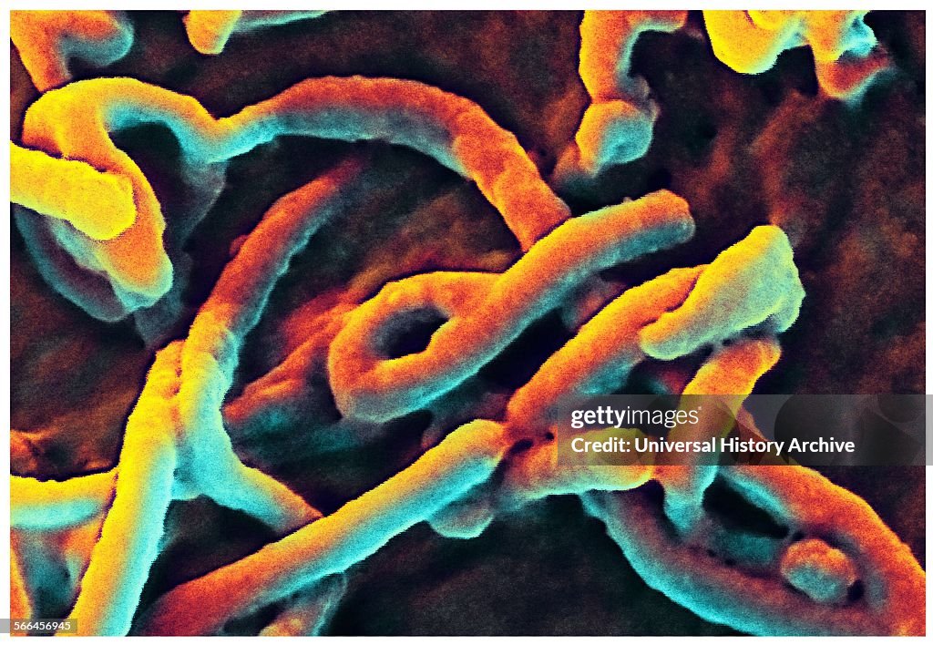
Scanning electron micrograph (SEM) depicts filamentous Ebola virus particles
Produced by the National Institute of Allergy and Infectious Diseases (NIAID), under a very-high magnification, this digitally-colourized scanning electron micrograph (SEM) depicts filamentous Ebola virus particles budding from the surface of a VERO cell of the African green monkey kidney epithelial cell line. (Photo by: Universal History Archive/Universal Images Group via Getty Images)

PURCHASE A LICENSE
How can I use this image?
kr. 2.200,00
DKK
Getty ImagesScanning electron micrograph (SEM) depicts filamentous Ebola virus..., News Photo Scanning electron micrograph (SEM) depicts filamentous Ebola virus... Get premium, high resolution news photos at Getty ImagesProduct #:566456945
 Scanning electron micrograph (SEM) depicts filamentous Ebola virus... Get premium, high resolution news photos at Getty ImagesProduct #:566456945
kr.3.000kr.850
Getty Images
In stockPlease note: images depicting historical events may contain themes, or have descriptions, that do not reflect current understanding. They are provided in a historical context. Learn more.
DETAILS
Restrictions:
Contact your local office for all commercial or promotional uses.
Credit:
Editorial #:
566456945
Collection:
Universal Images Group
Date created:
January 01, 1900
Upload date:
License type:
Release info:
Not released. More information
Source:
Universal Images Group Editorial
Object name:
917_05_WHA_057_0530
Max file size:
5100 x 3528 px (17.00 x 11.76 in) - 300 dpi - 3 MB